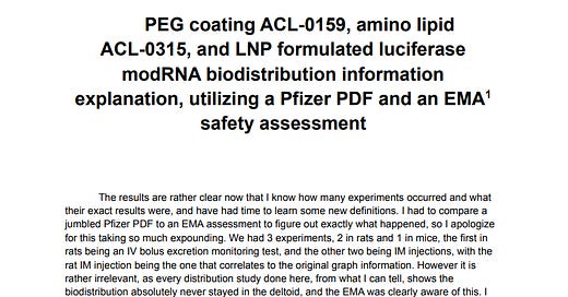
PEG coating ALC-0159, amino lipid ALC-0315, and LNP formulated luciferase modRNA biodistribution information explanation, utilizing a Pfizer PDF and an EMA safety assessment
I wrote this many months ago so I could try to figure out exactly where the lipid nanoparticles may distribute over what time frame, when injected either through intravenous or intramuscular pathways. It’s a personal assessment that has not been peer reviewed for error, so please be cautious and do your own due diligence on the work if you decide to use points from it. I had to interpret a Japanese version of the document combined with an EMA assessment, since I was having trouble locating the English PDF at the time, so sorry for any strange wording as a result. I have since found versions of both PDFs, copies below:
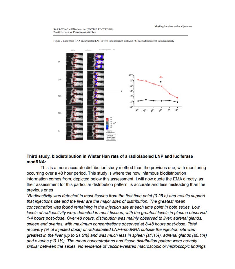
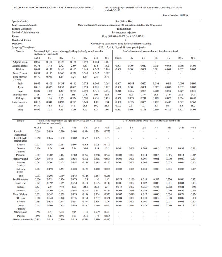
I don’t feel like messing with copy and pasting my assessment, so instead it will be included as pictures, with a PDF at the bottom:
AMENDMENT: Please disregard if you see “ALC” and “ACL” interchange in the PEG and lipid acronyms. The correct one, is “ALC”. As stated, the document may contain small irrelevant grammatical errors. Overall the assessment of the information however, I feel is very accurate.
As you can see, staying in the deltoid was never an option for the LNP platforms, and this was well known by governments that approved the shots. In fact I’d argue it should be obvious, as the coating of these LNP platforms (polyethylene glycol, or PEG https://en.wikipedia.org/wiki/Polyethylene_glycol )
is highly efficient at increasing the circulation half life of lipid nanoparticle carrier molecules. They have been studied for many decades, and this prolonging effect was always known, as the following excerpt from a paper dating back to 1990 shows:
”The activities of PEG-PE, Myrj 59 and GM1 in prolonging the circulation
half-life of the liposomes can also be seen in Fig. 2. The activity of PEG-PE was striking. At 1 h after injection, 85% of the injected dose was found in the blood and only 18% in the liver and spleen. Even at 5 h, there were still more liposomes in the blood (49%) than in the RES (38Ve). Myrj 59 also showed some activity although it was not stronger than that of G&Ii. The estimated fr/t for liposome blood clearance is ~30 min, 0.5 h, 1.5 h and 5 h for PC/chol liposomes and liposomes additionally cont~ning Myrj 59, GMr and PEG-PE, respectively. Thus, both amphipathic PEG significantly enhanced the blood residence time of the liposomes, although the activity of PEG-PE was superior to that of Myrj 59”
If mistakes are found, as usual, feel free to state where in the comments below


